Figure 1.
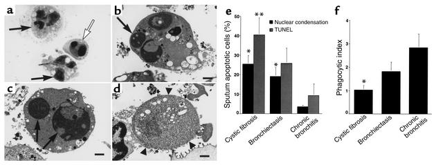
Clearance of apoptotic cells is defective in CF airways. (a) Wright’s Giemsa stain of CF sputum (×100) showing apoptotic cells with condensed nuclei (open arrow) and normal PMNs (arrows). (b–d) Transmission electron micrograph of sputum from a CF patient (×6,300; bars: 1 μm) demonstrating early apoptosis (b), late apoptosis (c), and postapoptotic necrosis (d). Arrows (b and c) indicate condensed apoptotic nuclei. Arrowheads (d) show loss of membrane integrity during necrosis. (e) Sputa from CF or bronchiectasis patients contain more apoptotic cells compared with chronic bronchitis. The percentage of apoptotic cells (by nuclear condensation and TUNEL staining) ± SEM is shown for six patients per group. *Apoptosis by nuclear condensation is significantly different from chronic bronchitis (P < 0.05). **Apoptosis by TUNEL staining is significantly different from chronic bronchitis (P < 0.05). (f) Airway macrophages from CF patients ingest fewer apoptotic cells. The mean phagocytic index of sputum macrophages ± SEM is shown for six patients per group. *Phagocytic index is significantly different from chronic bronchitis (P < 0.05).

