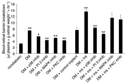
Figure 6.
Blood-retinal barrier breakdown in diabetic animals treated with insulin and kinase inhibitors. The diabetes-associated increases in blood-retinal barrier breakdown were suppressed with inhibitors of PKC, p42/p44 MAPK, p38 MAPK, or PI 3-kinase (7.45 to 4.37, 5.03, 5.4, and 4.25 μl plasma × g retinal dry weight–1 × hour–1, respectively). The insulin-associated increases in blood-retinal barrier breakdown were suppressed with the inhibition of p38 MAPK and PI 3-kinase (8.2 and 6.1 μl plasma × g retinal dry weight–1 × hour–1, respectively; n = 8, P < 0.001), whereas inhibition of PKC or p42/p44 MAPK had no effect (to 11.75 and 11.12 μl plasma × g retinal dry weight–1 × hour–1, respectively; n = 8, P > 0.5).

