
Fig. 3.
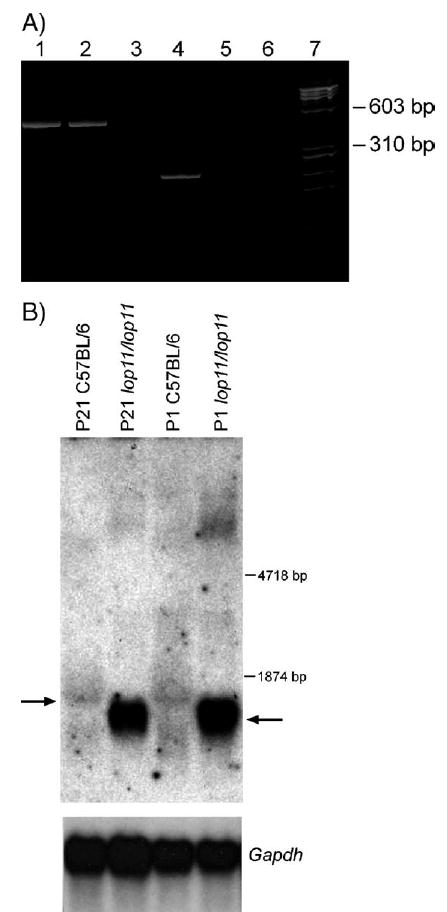
RT-PCR and Northern blot analysis. (A) Acrylamide gel electrophoresis of RT-PCR products derived from amplification of the Hsf4 gene from mouse eye mRNA. An RT-PCR product encompassing exons 1–6 is present in C57BL/6 and lop11/lop11 (lanes 1 and 2). An RT-PCR product encompassing exons 10–13 is present in C57BL/6, but absent from lop11/lop11 (lanes 4 and 5). Lanes 3 and 6 show H2O as a negative control; lane 7 is the φχ-174 RF DNA HaeIII molecular weight marker. (B) Northern analysis from P21 and P1 C57/BL6 and lop11/lop11 whole eye mRNA hybridized with a 5′ end Hsf4 probe (exons 1–6). Arrow to the left points to the wild-type Hsf4 transcript. Arrow to the right points to the lop11–Hsf4 transcript of the smaller molecular weight and higher level of expression. Hybridization of the same blot with Gapdh shows even loading between samples.