
Fig. 3.
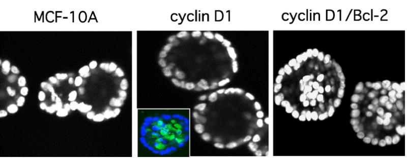
Cooperation between proliferative and survival signals results in filling of acinar lumen. Confocal sections of DAPI-stained day 20 structures formed by parental MCF-10As or cells overexpressing cyclin D1 or cyclin D1/Bcl-2 are shown. Inset, staining of cyclin D1 acini with DAPI (blue) and an antibody to the cleaved form of caspase-3 (green) reveals elevated levels of luminal apoptosis in these structures (13). The MCF-10A and cyclin D1 panels were reproduced from ref. (13).