Figure 3.

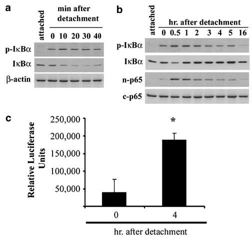
Detachment induces the activation of NF-κB. (a) Phosphorylated IκBα becomes detectable as a result of detachment and total protein levels decline over the first 30 min. p-IκB = phosphorylated-IκBα. (b) The protracted time course reveals that protein levels of IκBα recover by about 1 h following detachment. Secondly, increasing amounts of p65 protein accumulate in the nucleus coincidental with increased phosphorylated IκBα. n-p65, nuclear p65; c-p65, cytoplasmic p65. (c) IEC-18 were infected with adenovirus expressing NF-κB-promoted luciferase prior to detachment, then detached and harvested immediately or after 4 h, and luciferase activity was measured. *P < 0.01
