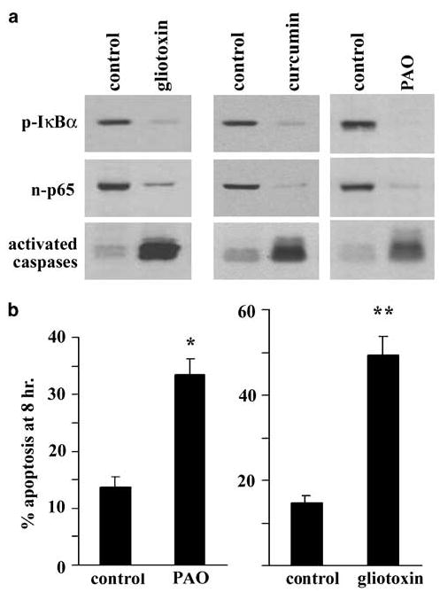
Figure 5.

(a) Pharmacological inhibitors of NF-κB prevent phosphorylation of IκBα and the nuclear accumulation of p65 (assayed at 1 h) as well as accelerate the activation of caspases, detected with the pan activated caspase marker zEK (bio) D-aomk using 4 h lysates. Cells were incubated in each inhibitor during trypsinization and throughout the period of detachment until harvest. The blots were stripped and probed for actin, which showed similar levels of protein in each lane (not shown). (b) The proapoptotic effect of gliotoxin and PAO quantitated using the morphology of detached cells after 8 h. Gliotoxin was used at 0.5 μg/ml, curcumin at 50 μm and PAO at 0.5 μm. *P < 0.05, **P < 0.01
