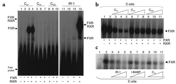
Figure 8.
FXR binds the human apoA-I promoter C site. (a) Gel retardation assays were performed on end-labeled Cwt (lanes 1–4), Cmt (lanes 5–7), CGG (lanes 8–10), and IR-1 consensus FXRE (lanes 11–14) oligonucleotides in the presence of in vitro–transcribed/translated unprogrammed reticulocyte lysate (lanes 1 and 11), mRXR (lanes 2, 5, 8, and 12), hFXR (lanes 3, 6, 9, and 13), or both mRXR and hFXR (lanes 4, 7, 10, and 14). (b) Gel retardation assays were performed on end-labeled wild-type C-site oligonucleotide in the presence of in vitro–transcribed/translated hFXR and mRXR. Competitions were performed by adding 100-fold (lanes 3, 6, and 9), 200-fold (lanes 4, 7, and 10), or 400-fold (lanes 5, 8, and 11) molar excess of cold Cwt wild-type (lanes 3–5), Cmt (lanes 6–8), or CGG (lanes 9–11) oligonucleotides, respectively. (c) Gel retardation assays were performed on end-labeled wild-type C-site oligonucleotide in the presence of in vitro–transcribed/translated hFXR and mRXR proteins. Competitions were performed by adding tenfold (lanes 2, 5, and 8), 50-fold (lanes 3, 6, and 9), or 100-fold (lanes 4, 7, and 10) molar excess of cold IR-1 consensus FXRE (lanes 2–4), I-BABP (lanes 5–7), or Cwt wild-type (lanes 8–10) oligonucleotides, respectively.

