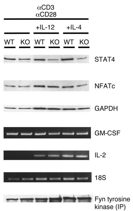
Figure 7.
Activation of pathways involved in T cell activation and differentiation in spleen cells from VDR KO and WT mice. Spleen cells were stimulated with 10 μg/ml anti-CD3 and 2 μg/ml anti-CD28 for 3 days, with either 100 ng/ml IL-12 and 50 μg/ml anti–IL-4 or 10 ng/ml IL-4 and 10 μg/ml anti–IL-12. Protein expression of STAT4, NFATc, and GAPDH was determined by Western blot analysis after 3 days of stimulation. Immunoprecipitation (IP) of protein lysates was performed with anti-fyn antibody, and protein A–precipitated product was probed with an antibody against tyrosine kinase to detect phosphorylation. Ethidium bromide gels show RT-PCR for GM-CSF, IL-2, and 18S after stimulation for 1 day.

