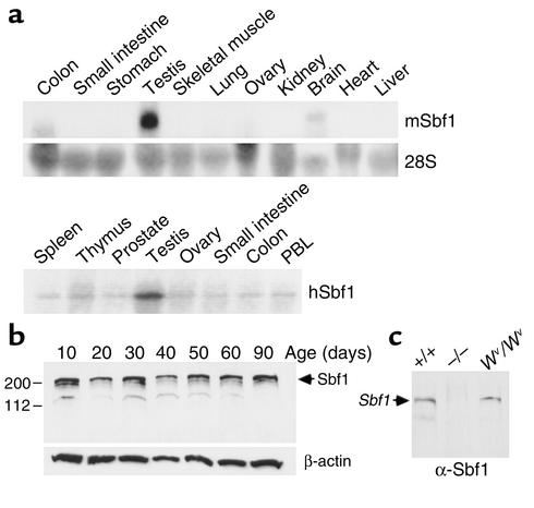
Figure 1.
Tissue-specific expression of Sbf1. (a) Northern blot analysis of total RNA prepared from human tissues or adult mice. The hybridization probe consisted of an Sbf1 cDNA (upper panel). Abundance of 28S rRNA is shown for the mouse blot for comparison. (b) Western blot analysis of protein extracts from testes at different stages of spermatogenic development. Migration of Sbf1 (upper panel) and β-actin (loading control, lower panel) is indicated. Faint bands in some lanes at 150 kDa represent apparent Sbf1 degradation products. (c) Anti-Sbf1 Western blot analysis of protein extracts from wild-type, Sbf1–/–, and Wv/Wv adult mice.

