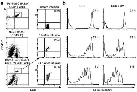
Figure 5.
Activation of donor naive CD8+ T cells upon intravenous injection into lethally irradiated B6 recipients. (a) Naive CD8+CD45.2+ C3H.SW T cells (2 × 10⁶) were intravenously injected into lethally irradiated B6/SJL recipients (CD45.1+). At the indicated timepoints, splenocytes were prepared from these mice and tricolor-stained with anti-CD8, anti-CD45.2, and anti-CD25 Ab’s, which were separately revealed with FITC, Cychrome, and PE. After gating on CD45.2+ donor–derived T cells, the expression of CD25 on CD8+ T cells was analyzed by flow cytometry. (b) CFSE-labeled CD8+ T cells (2 × 10⁶) from C3H.SW donor mice (CD45.2) were transferred with or without mixed donor T–BM cells into lethally irradiated B6/SJL recipient mice (CD45.1). At the indicated timepoints after transplantation, splenocytes were prepared from these mice, stained with anti-CD8 Ab conjugated with Cychrome, and then sequentially labeled with biotinylated anti-CD45.2 and streptavidin-conjugated PE. The division of donor CD8+ T cells in vivo was analyzed by flow cytometry by gating on CD8+CD45.2+ T cells. One representative experiment of three is shown.

