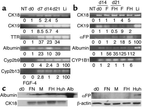
Figure 4.
Quantitative RT-PCR and Western blot analyses confirm hepatocyte-like phenotype. (a) mMAPCs and (b) hMAPCs were cultured on Matrigel with FGF-4 and HGF or FGF-4 alone for 21 and 28 days, respectively. At multiple time points during culture, cells were harvested and underwent quantitative RT-PCR using the SYBR green method for mRNAs as indicated. The mRNA levels were normalized using β-actin as a housekeeping gene. Numbers under the blots for CK18, CK19, TTR, albumin, and CYP1B1 represent mRNA levels after treatment relative to undifferentiated MAPCs. For αFP, Cyp2b9, and Cyp2b13, numbers under the blots are relative to mRNA from the liver, because no transcripts were detected in undifferentiated MAPCs. Li, mouse or human liver mRNA; NT, no template. Representative example of five mouse and one human study. (c) hMAPCs (from b) were cultured on Matrigel with FGF-4 and HGF or FGF-4 alone for 21 days. HUH7 cells were cultured as described. Lysates of cells were separated by SDS-PAGE, transferred to Immuno-Blot PVDF membrane, and incubated for 1 minute with Ab’s against αFP, CK18, albumin, or β-actin. FN, FGF-4–induced hMAPCs on FN; M, FGF-4–induced hMAPCs on Matrigel; FH, FGF-4– and HGF-induced hMAPCs on Matrigel; Huh, Huh7 cell line used as control.

