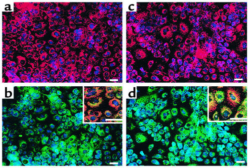
Figure 7.
LDL uptake by hMAPC-derived hepatocytes. Thee hMAPCs cultured with FGF-4 on Matrigel for 0–21 days were incubated with Dil-acil-LDL, fixed, and stained with anti–pan-CK, CK18, GATA4, and HNF-3β Ab’s. Representative example of three studies. Scale bar, 25 μm. (a) Day 21, incubated with Dil-acil-LDL and stained with HNF-3β-Cy5 Ab. (b) Same field as a stained with pan-CK-FITC and HNF-3β-Cy5 Ab. Inset, higher-magnification three-color view. Note the presence of binucleated cells. (c) Day 21, incubated with Dil-acil-LDL and stained with GATA4-Cy5 Ab. (d) Same field as b stained with GATA4-Cy5 and CK18-FITC Ab. Inset, higher magnification showing three-color view. Note the presence of binucleated cells.

