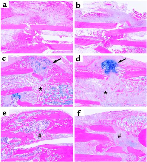
Figure 2.
Histologic evidence of defective fracture healing in COX-2–/– mice. Histologic sections were obtained from tibial fractures of adult wild-type (a, c, and e) and COX-2–/– mice (b, d, and f) at 7 (a and b), 14, (c and d), and 21 days (e and f) after fracture, and stained with Alcian blue/hematoxylin as described in Methods. Fractures in wild-type mice undergo extensive calcification resulting in little residual cartilage (arrow) and extensive woven bone formation (asterisk) by day 14 (c). This progresses to a remodeling bony union (#) by day 21. In the COX-2–/– mice there is little evidence of endochondral bone formation in the medullary canal, which is filled with undifferentiated mesenchymal tissue (asterisk) on day 14 (d). Concurrently, significant amounts of unmineralized cartilage persists (arrow) (d). By 21 days there are large amounts of fibrotic tissue (#) between the fractured bones, evidence of a fracture nonunion (f).

