Figure 5.
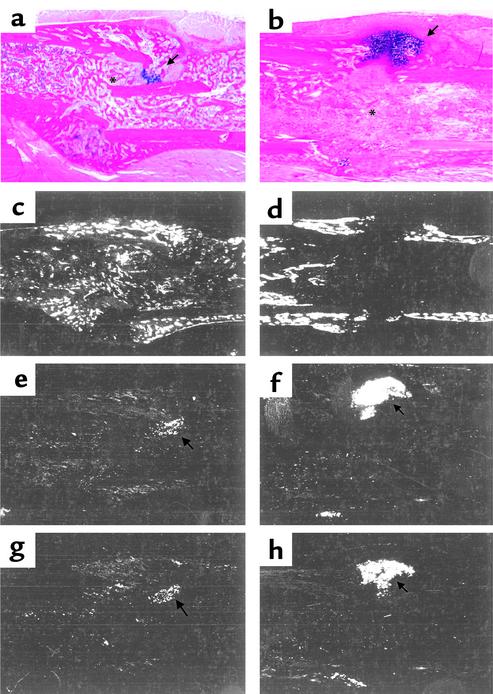
Characterization of defective fracture healing in COX-2–/– mice by in situ hybridization. Histologic sections of the fracture callus of wild-type (a, c, e, and g) and COX-2–/– mice (b, d, f, and h), 14 days after fracture, were stained with Alcian blue/hematoxylin (a and b). Serial sections were used for in situ hybridization with probes specific for osteocalcin (c and d), col2 (e and f), and colX (g and h). The gene expression profile confirms the persistence of cartilage (arrows) and the decrease in osteogenesis (asterisks) in COX-2–/– mice.

