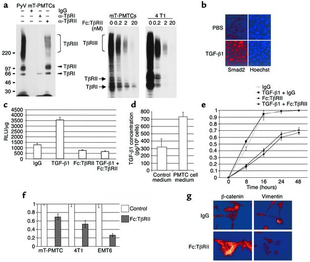
Figure 2.
Fc:TβRII inhibits autocrine TGF-β signaling in PyV mT PMTCs. (a) Left panel: PMTCs were affinity labeled with 125I-TGF-β1, resolved directly (lane 1) or immunoprecipitated with the indicated TβR antibodies or IgG. Right panel: PMTCs and 4T1 cells were labeled with 125I-TGFβ1 in the presence of the indicated concentrations of Fc:TβRII. (b) Immunofluorescent detection of Smad2 in PMTCs treated with PBS or TGF-β1. (c) PMTCs transfected with a 3TP-Lux were treated with Fc:TβRII, TGF-β1, or both. The results are presented as relative light units (RLUs)/μg of total protein.The values represent the average of 3 experiments performed in duplicate, ± SE. (d) PMTCs or PMECs were cultured in serum-free medium for 24 hours. Conditioned medium was collected and analyzed for TGF-β1 using ELISA. Results are shown as an average of 5 experiments, analyzed in triplicate. (e) PMTCs were grown to confluency and wounded. The results are presented as the percentage of the total area of the original wound enclosed by cells and represent the average ± SD obtained from five experiments, analyzed in triplicate. (f) Transwell assays were performed using PMTCs, 4T1 cells, or EMT6 cells. The number of cells migrating to the lower side of the filter in controls was given the value of 1, such that migration of cells is represented as a fraction of control. Values shown are the average (± SE) of triplicate transwells in three experiments. (g) Immunocytochemical detection of β-catenin or vimentin in PMTCs cultured in the presence of the indicated factors. Representative photographs are shown.

