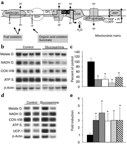
Figure 2.
In vivo effect of GlcN on the expression of nuclear-encoded mitochondrial transcripts. (a) Schematic illustration of respiratory chain complexes and oxidative phosphorylation. Electrons (e–) donated from fuel oxidation are sequentially transferred from ubiquinone (Q and QH2) to cytochrome c (Cyt. c), and finally to oxygen (O2). Simultaneously, protons (H+) are translocated across the inner mitochondrial membrane and create an electrochemical gradient whose energy is eventually used by ATP synthase for ATP formation. An adenine nucleotide translocator (ANT) carries ATP out of the mitochondria in exchange for ADP. The complexes indicated in gray contain one or more subunits downregulated by glucosamine. COX, cytochrome c oxidase; SDH, succinate dehydrogenase. (b) Northern analysis of mitochondrial transcripts in skeletal muscle from glucosamine versus saline infusion (control) with probes encoding mitochondrial malate dehydrogenase, NADH dehydrogenase, cytochrome c oxidase (COX-VIB), ATP synthase, and β-actin. (c) Quantitation, by densitometry, of Northern blots shown in b. Data are expressed as percent of control hybridization and are normalized with a β-actin probe. (d) Northern analysis of mitochondrial transcripts in BAT from glucosamine versus saline infusion (control) studies. Probes are the same as in b, except for the BAT-selective UCP-1. (e) Quantitation of Northern blot shown in d. Legend to bar graphs: Black, control; dark gray, UCP-1; medium gray, ATP synthase; white, cytochrome c oxidase VIB; light gray, NADH dehydrogenase; hatched, malate dehydrogenase. *P < 0.01.

