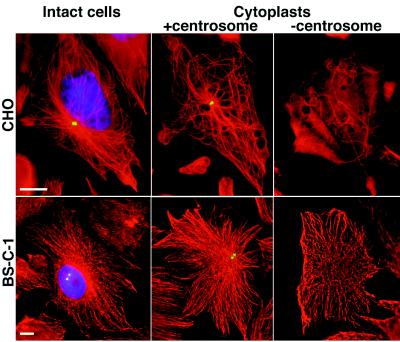
Figure 1.
Distribution of MTs in intact cells and cytoplasts. Intact cells (Left) and cytoplasts containing (Center) or lacking (Right) the centrosome were fixed and triple-stained with α-tubulin antibody for MTs (red), γ-tubulin antibody for the centrosome (green, but superposition makes the spot appear yellow) and 4′,6-diamidino-2-phenylindole (DAPI) for the nucleus (blue). (Upper) CHO cells; (Lower) BSC-1 cells. (Bar = 10 μm.)

