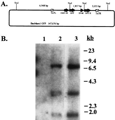
Figure 7.
Analysis of baculovirus-derived DNA in stably transduced CHO cells. (A) Map of BacMam1 GFP recombinant baculovirus DNA. StuI recognition sites internal and adjacent to the recombinant expression cassettes are shown with sizes of the expected fragments. Drawing is not to scale. (B) Southern blot of StuI-digested DNA. Details are described in the text. Lanes: 1, nontransduced cells; 2, transduced cells, selected pool; 3, BacMam1 GFP viral DNA.

