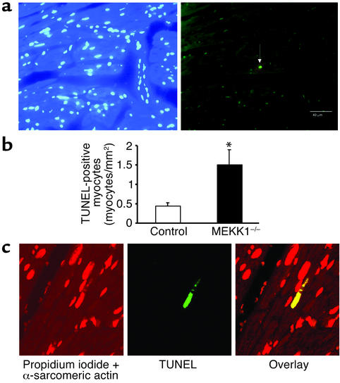
Figure 4.
Pressure overload causes more TUNEL-positive cells in the left ventricle in MEKK1–/– mice. (a and b) DAPI staining (left) and TUNEL staining (right) of the LV myocardium 7 days after aortic banding in a MEKK1–/– mouse. The white arrow indicates a TUNEL-positive nucleus. Bar = 40 μm. (b) TUNEL-positive myocytes in LV myocardium were counted in control and MEKK1–/– mice subjected to aortic banding for 7 days and expressed as the number per mm2. The number of TUNEL-positive myocytes was significantly higher in MEKK1–/– mice than in the control mice. *P < 0.05; n = 5. (c) Images of the confocal microscopic analyses showing nuclear fragmentation in a MEKK1–/– mouse banded for 7 days. Triple staining (propidium iodide, TUNEL, and anti–α-sarcomeric actin antibody) was performed. Staining for propidium iodide and anti–α-sarcomeric actin antibody is shown by red, and that for TUNEL by green. In the overlay image, a nucleus stained by both TUNEL and propidium iodide is shown by yellow.

