Figure 5.
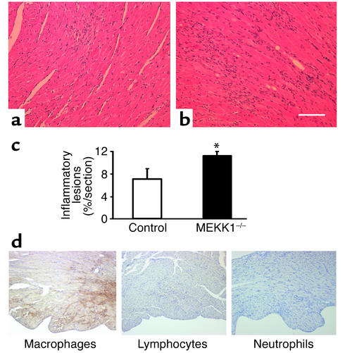
Pressure overload causes more inflammatory lesions, predominantly consisting of macrophages, as well as apoptosis of the inflammatory cells in the left ventricle in MEKK1–/– mice. Hematoxylin-and-eosin staining reveals an increased number of inflammatory lesions in MEKK1–/– mice (b) compared with control mice (a). Bar = 150 μm.(c) The inflammatory lesions in LV myocardium were measured by image analysis software. There was an increase in inflammatory lesions in MEKK1–/– mice compared with control mice after 7 days of pressure overload. *P < 0.05; n = 5. (d) Macrophages, lymphocytes, and neutrophils in the LV myocardium from MEKK1–/– mice subjected to pressure overload were stained as described in Methods. The inflammatory cells primarily consisted of macrophages.

