
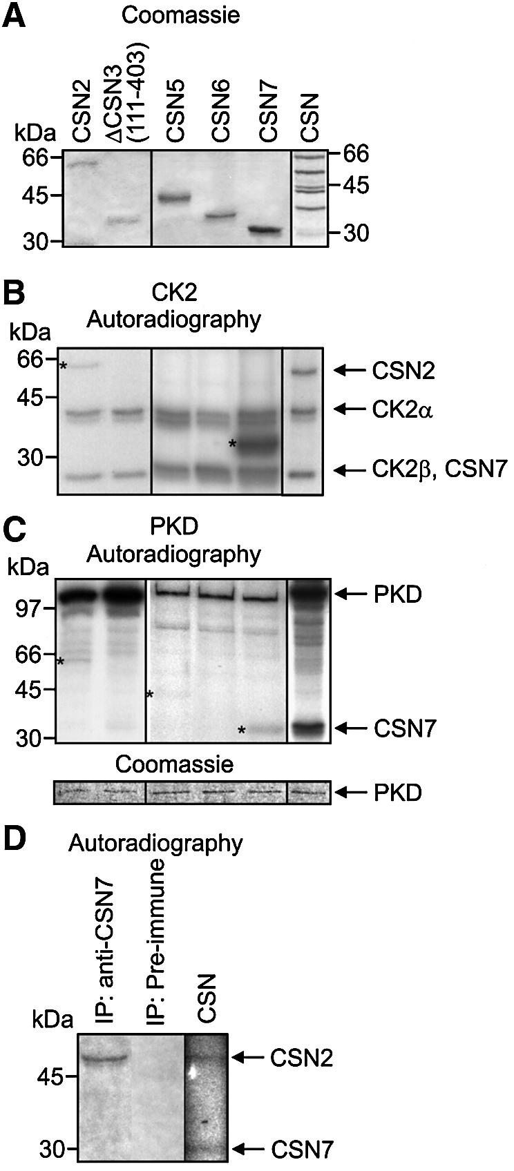
Fig. 4. CK2 and PKD phosphorylate subunits of the CSN. (A) In vitro kinase assays were performed with shown recombinant CSN subunits and purified CSN as substrates (Coomassie). (B) Each recombinant CSN subunit or purified CSN were incubated with recombinant CK2. Autoradiography shows phosphorylated recombinant CSN subunits labeled with stars. Autoradiography of the phosphorylated CSN complex reveals that phosphorylated CSN7 and autophosphorylated CK2β co-migrate in SDS–PAGE. Complex-bound subunits migrate faster in SDS–PAGE as compared with recombinant His6-tagged CSN subunits. (C) Kinase reactions were carried out as in (B) using recombinant PKD. Phosphorylated recombinant subunits are indicated by stars. Different proteins exert different effects on autophosphorylation of recombinant kinases. To demonstrate that equal amounts of PKD were added to each sample, Coomassie Blue-stained PKD is shown (lower panel). (D) Subunit CSN2 of endogenous CSN is phosphorylated in reticulocyte lysate. Reticulocyte lysate was incubated with [γ-32P]ATP. After incubation endogenous CSN was immunoprecipitated and the precipitate was analyzed by SDS–PAGE and autoradiography. Control (CSN): purified CSN was incubated with [γ-32P]ATP and then analyzed by SDS–PAGE and autoradiography.