

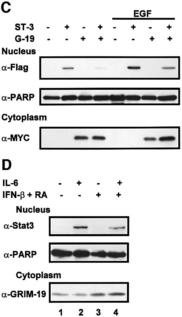
Fig. 6. Co-localization of Stat3 with GRIM-19. (A) MCF-7 cells were transfected with Flag-tagged Stat3 or truncation mutants with or without Myc-tagged GRIM-19. The distribution of Stat3 or GRIM-19 was detected using monoclonal anti-Flag or polyclonal anti-Myc as primary antibody, followed by Cy3-conjugated anti-mouse IgG (Sigma) or FITC-conjugated anti-rabbit IgG. (B) COS-1 cell were transfected with Flag-tagged Stat3, Stat1 or Stat5a in the absence (–) or presence of Myc-GRIM-19. The cells were induced by EGF for 15 min and the cellular localization of Stat proteins and GRIM-19 was detected as described in (A). (C) The effect of GRIM-19 on Stat3 nuclear translocation. COS-1 cells were transfected with vector, GRIM-19 and/or Stat3. The cells were untreated or induced with EGF for 15 min. Cells were lysed and fractionated into cytoplasmic and nuclear portions as described previously (Jain et al., 1999). The nuclear distribution of Stat3 was examined by western blotting. The blot was re-blotted with anti-PARP antibody as a control. The expression of GRIM-19 in the cytoplasmic portion was detected by blotting with anti-Myc antibody. (D) Induction of endogenous GRIM-19 reduced IL-6-stimulated Stat3 nuclear translocation. HepG2 cells were either left untreated or treated with IFN-β and RA for 48 h followed by IL-6 induction for 15 min. Nuclear Stat3 was measured with anti-Stat3 as described in (C).