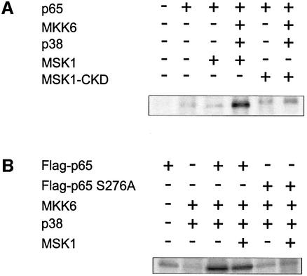
Fig. 5. MSK1 phosphorylates p65 in vivo. (A) HEK293 cells, transiently transfected with the relevant expression vectors, were labeled with [32P]orthophosphate for 4 h. Cell lysates were subjected to immunoprecipitation with anti-p65 antibody. Precipitated proteins were separated by SDS–PAGE and analyzed using PhosphorImager technology. (B) Similar experiment to that in (A), but immunoprecipitation was performed with anti-Flag antibody.

An official website of the United States government
Here's how you know
Official websites use .gov
A
.gov website belongs to an official
government organization in the United States.
Secure .gov websites use HTTPS
A lock (
) or https:// means you've safely
connected to the .gov website. Share sensitive
information only on official, secure websites.
