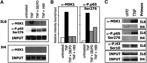
Fig. 7. MSK1 and its phosphorylated substrates position at the endogenous IL-6 promoter upon TNF treatment. L929sA mouse fibroblasts were starved for 24–48 h in 0.5% serum. Quiescent cells were treated for 30 min with 2500 IU/ml TNF alone, or following 2 h pre-treatment with the inhibitors SB203580 + PD98059 (10 µM) or H89 (10 µM). ChIP analysis was performed against MSK1, or against phospho NF-κB p65 (Ser276) (A and C) or phospho H3 (Ser10) (C). After reversal of cross-linking, co-immunoprecipitated genomic DNA fragments were analyzed by quantitative PCR for 27 cycles with IL-6 or H4 promoter-specific primer sets. Input reflects the relative amounts of sonicated DNA fragments present before immunoprecipitation and revealed by quantitative PCR with either IL-6- or H4-specific primers. (B) Schematic representation of the results obtained in (A).

An official website of the United States government
Here's how you know
Official websites use .gov
A
.gov website belongs to an official
government organization in the United States.
Secure .gov websites use HTTPS
A lock (
) or https:// means you've safely
connected to the .gov website. Share sensitive
information only on official, secure websites.
