Figure 7.
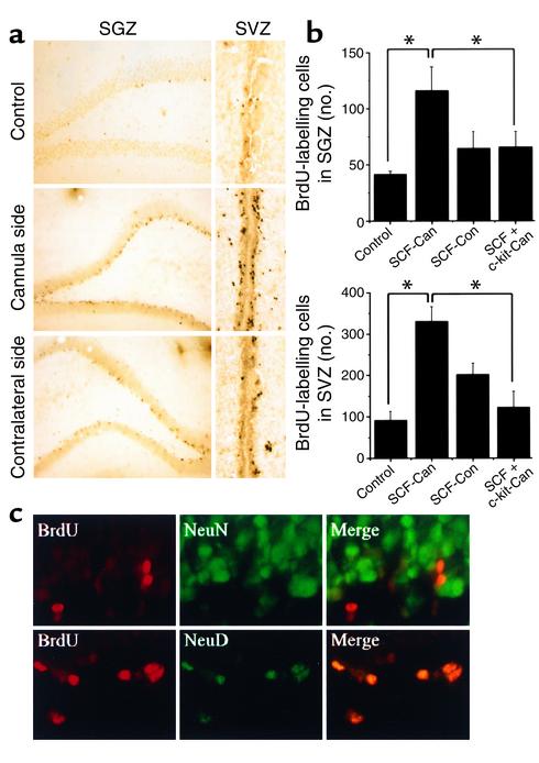
Intraventricular SCF stimulates BrdU incorporation in SGZ and SVZ in vivo. (a) Brain sections through SGZ and SVZ were immunostained with anti-BrdU Ab 1 week after intraventricular infusion of SCF or aCSF vehicle (n = 6). Compared with aCSF (control), SCF increased the number of BrdU-positive cells in SGZ and SVZ. Proliferation was more pronounced on the cannula side, as compared with the contralateral side. (b) BrdU-labeled cells in SGZ (top) and SVZ (bottom) were counted in control brain (n = 6) and both ipsilateral (cannula side) and contralateral to SCF infusion (n = 6). In some experiments, SCF was infused together with anti–c-kit Ab (n = 4). Bars (left to right) represent control; SCF-treated, cannula side; SCF-treated, control side; and SCF- and anti–c-kit-treated, cannula side. BrdU-positive cells were increased in SVZ on both the infused and contralateral sides and in SGZ on the infused side.The effect of SCF was partially blocked by coadministration of anti–c-kit Ab (*P < 0.05, Student t test). (c) Rat brain sections through SGZ, obtained 1 week after SCF infusion, were double-labeled for BrdU (red) and NeuN or Neuro D (green). Merged images show that BrdU labeling colocalized with Neuro D and, in some cases, NeuN. Data shown are representative fields from the number of experiments given above (a and c), or mean ± SEM (n = 3) (b).

