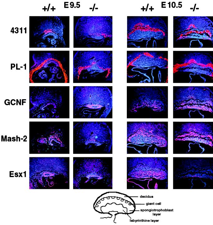
Figure 4.
Expression of cell-type-specific markers analyzed by in situ hybridization of sagittal sections of conceptus at E9.5, and placentas from Dlx3 +/+ and −/− embryos at E10.5. Hybridizations were performed with antisense probes for 4311 (spongiotrophoblast cells and their precursors in the ectoplacental cone); PL-1 (trophoblast giant cell); mGCNF (labyrinthine layer); Mash-2 (spongiotrophoblast and labyrinthine layers and chorion); and Esx1 (endoderm layer of the visceral yolk sac and labyrinthine trophoblast of the chorioallantoic placenta). (×10.) At the bottom of the figure there is a schematic representation of E10.5 placenta indicating the different cell types.

