Figure 4.
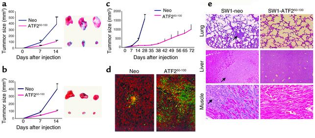
(a) Mouse melanoma expressing the ATF250–100 peptide grow to a substantially smaller size. SW1 cells that express control vector or the ATF250–100 peptide were injected subcutaneously into C3H/HEJ mice, and tumor growth was monitored. The right panel depicts representative tumors at the end of the study, and the left panel shows tumor growth. The experiment was repeated three times. Bars = SD; P < 0.0001. (b) SW1 cells that express empty vector or the ATF250–100 peptide were injected subcutaneously into C3H/GLD (GLD mice bear mutations in FasL) mice, and follow-up and analysis were performed as indicated in a. Bars = SD; P < 0.0001. (c) Long-term growth of tumors from ATF250–100 peptide–expressing SW1 cells in C3H/HEJ mice. Tumors were measured at the indicated time points. Bars = SD; P < 0.0003. (d) Apoptosis of SW1 tumors expressing the ATF250–100 peptide. Sections of SW1 tumors produced in the presence of ATF250–100 peptide or control vector, tested for apoptosis by the TUNEL assay (apoptotic cells are green). (e) Expression of the ATF250–100 peptide in SW1 cells inhibits metastasis. Mice bearing tumors produced by cells that constitutively express the ATF250–100 peptide were monitored for metastatic lesions in the indicated organs at the end of inoculation. Arrows point to the metastatic lesions seen in the control group at the end of their inoculation.

