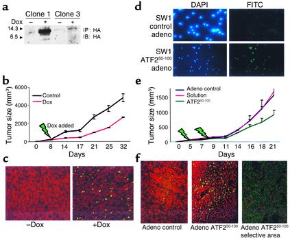
Figure 5.
Administration of ATF250–100 peptide to existing SW1 tumors reduces their growth in C3H mice. (a–c) SW1 cells that exhibit inducible expression of the ATF250–100 peptide following addition of doxycycline were selected (a) and injected (clone 1) into C3H mice. Eight days after inoculation, doxycycline was added to the drinking water. Tumors were measured at the indicated times (b; P < 0.003). Analysis of apoptosis via the TUNEL assay revealed marked apoptosis in the tumors obtained from doxycycline-treated mice (c, green staining). (d) SW1 cells were infected with adenovirus (adeno) control or with adenovirus that expresses ATF250–100 peptide, and then subjected to immunohistochemistry analysis using antibodies to HA (HA is cloned in frame with the ATF2 peptide sequence). Left panels depict DAPI staining, whereas right panels show the fluorescence upon positive staining with HA antibodies. (e) SW1 cells were injected subcutaneously and tumors reached the size of 40 mm3 before virus control, solution control, or virus carrying the ATF2 peptide was injected (green arrows indicate injection). Data shown represent two experiments. P < 0.02 between control virus and the ATF2 peptide adenovirus groups. (f) Analysis of tumors for apoptosis via the TUNEL assay. Comparable magnification of control virus and ATF2 virus is shown in the two left panels; the right panel depicts a selective area representative of marked apoptosis within focal areas, seen in tumors that express the ATF2 peptide.

