
Figure 5.
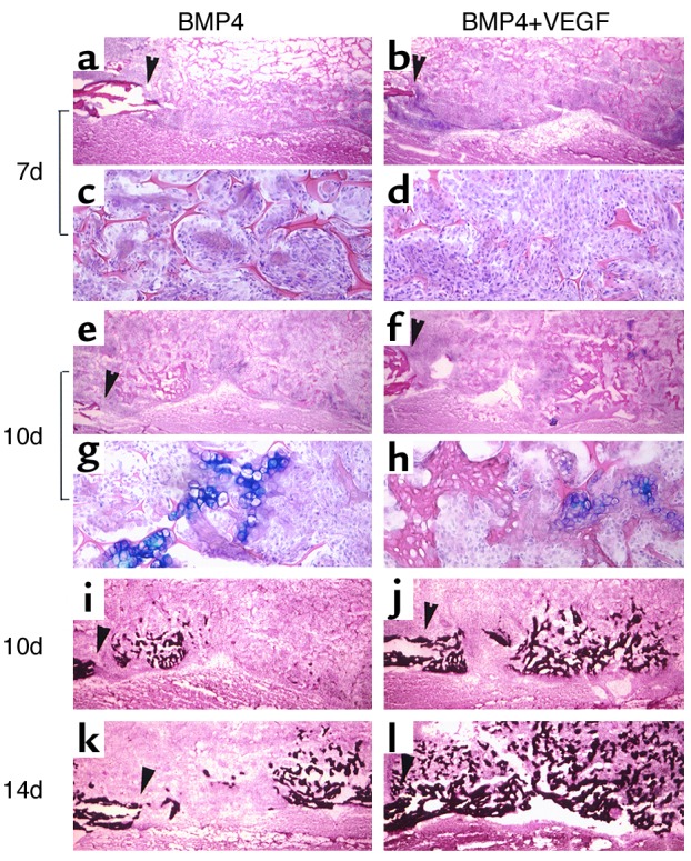
Critical-sized calvarial defects healed through the endochondral ossification pathway in both BMP4 and BMP4+VEGF groups. (a–h) Alcian Blue staining shows cartilage formation in the defects implanted with transduced MDSCs. The edges of the bone defects are marked by arrowheads. Mesenchymal cell infiltration was more abundant in the BMP4+VEGF group (b and d) than in the BMP4 group (a and c) at 7 days PI. c and d display local magnifications of a and b, respectively. At 10 days, cartilage bridging the defects was mostly resorbed, leaving traces of hypertrophic chondrocytes (e–h). g and h display local magnifications of e and f, respectively. (i–l) von Kossa staining demonstrates coupled bone mineralization at 10 days and increased mineralized bone formation at 14 days in the BMP4+VEGF group (j and l) compared with the BMP4 group (i and k). Magnification: a, b, e, f, and i–l, ×40; c, d, g, and h, ×200.