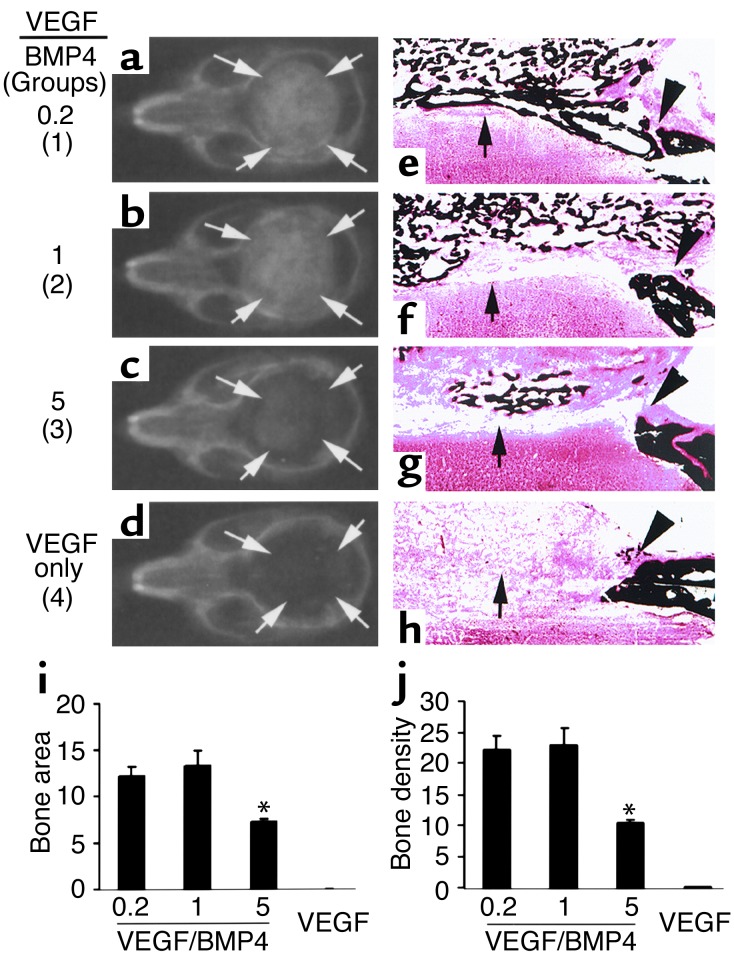
Figure 7.
The proper ratio of VEGF to BMP4 is critical to ensure a beneficial effect on bone healing. In groups 1–3, the ratios of VEGF- to BMP4-expressing cells were 0.2, 1, and 5, respectively; in group 4, only VEGF-expressing cells were used. Arrows mark the edge of the bone defects and regenerated bone. (a–d) Radiography shows complete bone healing in groups 1 and 2, partial bone healing in group 3, and no bone healing in group 4. (e–h) von Kossa staining shows regeneration of cortical bone–like structure (arrow) in group 1 (e) but not in other groups, although bone union also occurred in group 2 (f). Bone regeneration was significantly reduced in group 3 as compared with groups 1 and 2 (e–g). Only fibrous tissue fills the defect in group 4 (h). Arrowheads mark the edge of the critical-sized bone defects and the regenerated bone. Magnification: ×40. (i and j) Quantitative analysis shows a significant decrease in bone regeneration in group 3 as compared with groups 1 and 2, while no bone formation was detected in the group containing only VEGF-expressing cells. Group 1 vs. group 2, P > 0.05, n = 4; group 1 vs. group 3, P < 0.01, n = 4; *P < 0.01 when compared to group 1.

