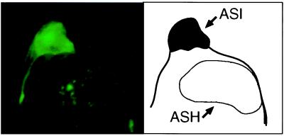
Figure 2.
Htn-Q150/OSM-10∷GFP expression led to morphological changes of the ASH in aged animals. (Left) An Htn-Q150/ OSM-10∷GFP-expressing ASH neuron from an 8-day-old animal with a speckled bag phenotype, visualized by GFP fluorescence. Multiple serial section confocal planes were combined for this image. Htn-Q150/OSM-10∷GFP expression does not affect the size and morphology of the ASI sensory neurons (as illustrated), and the GFP marker in the ASI neuron shows the normal subcellular expression pattern (A.C.H., J. Kaas, J. E. Shapiro, and J. M. Kaplan, unpublished work). In contrast, a subset of ASH sensory neurons are severely affected, displaying a speckled bag phenotype, swelling to 2–3 times the size of a normal ASH neuron, and lacking intracellular morphology. The subcellular structure, if any, that contains the faint GFP-positive dots is unclear. The size of the ASH and ASI sensory neurons are comparable in wild-type animals. osm-10 expression in ASH neurons is significantly higher than expression in ASI neurons (A.C.H., J. Kaas, J. E. Shapiro, and J. M. Kaplan, unpublished work), and, consequently, polyQ induced effects are less common in ASI neurons. (Right) A schematic representation of the upper panel. The ASI and ASH neurons are outlined.

