Figure 3.
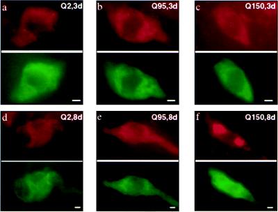
Htn-Q150 expression led to time-dependent protein aggregation in sensitized ASH neurons. (a–c Upper) Visualization of Htn-Q2, Htn-Q95, and Htn-Q150 expression in sensitized ASH sensory neurons in 3-day-old transgenic animals by using the antihuntingtin HP1 (35) antiserum in red. All huntingtin fragments were expressed at similar levels and were found in the cytoplasm and processes without accumulation in obvious intracellular structures. For Htn-Q150, the huntingtin fragments accumulated into discrete foci or aggregates in a very small percentage of the Htn-Q150 expressing ASH sensory neurons (aggregates were found in random locations in the cytoplasm and processes but not in the nucleus, based on visual examination). (a–c Lower) The ASH sensory neuron shown in the upper panels visualized with the GFP marker in green. (d–f Upper) Visualization of Htn-Q2, Htn-Q95, and Htn-Q150 expression in sensitized ASH sensory neurons in 8-day-old transgenic animals by using the antihuntingtin HP1 (35) antiserum in red. Localization of Htn-Q2 and Htn-Q95 fragments was still broadly cytoplasmic. For Htn-Q150, the huntingtin fragments were detected in large and small cytoplasmic aggregates in over half of the Htn-Q150-expressing ASH neurons. (d–f Lower) The ASH sensory neuron shown in the upper panels visualized with the GFP marker in green. (Bar = 3 μm.)

