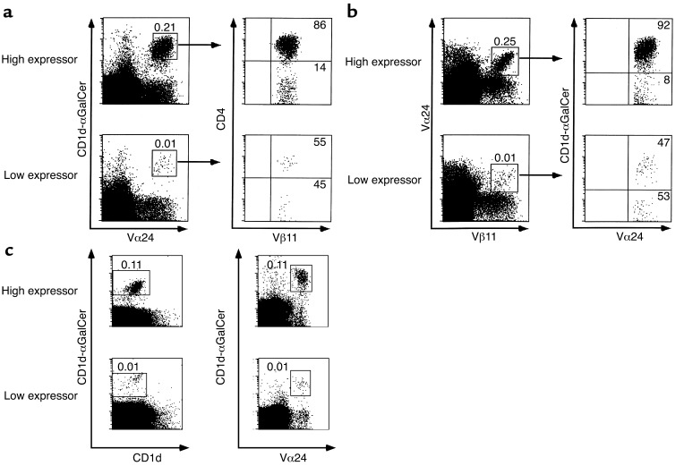
Figure 1.
Specific identification of human NKT cells in PBLs. (a) NKT cells in PBLs were identified by double staining with CD1d-αGalCer tetramers followed by anti-Vα24 mAb (left panels). The CD4 distribution and Vβ11 usage among these cells were identified in the same staining (right panels). (b) Vα24/Vβ11 double-positive PBLs (left panels) were stained with CD1d-αGalCer tetramers (right panels). Numbers above the boxes represent percentages of cells among total PBLs, whereas numbers in the quadrants represent percentages among boxed cells. Representative FACS plots of donors expressing high or low frequency of NKT cells are shown. (c) Double staining with CD1d-αGalCer and empty CD1d (left panels) identified the same percentage of NKT cells as double staining with CD1d-αGalCer and Vα24 (right panels).

