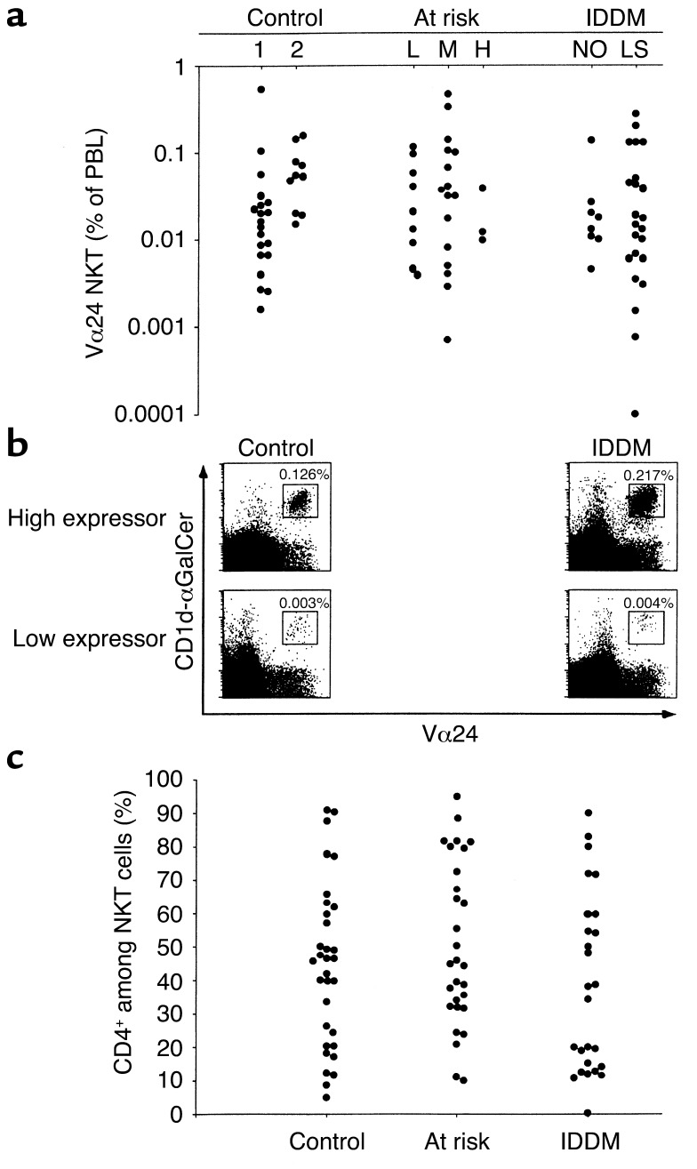
Figure 3.
Conserved expression of NKT cells in individuals with IDDM. (a) Summary plot showing individual results in controls, subdivided in age- and DQ-matched individuals (cohort 1) and healthy volunteers (cohort 2), at-risk individuals (separated into low [L], medium [M], and high [H] risk), and IDDM new onset (NO) and long-standing (LS) individuals. (b) Representative FACS plots of healthy or IDDM individuals expressing high or low frequency of NKT cells in PBLs. (c) Summary plot of the relative proportion of CD4+ cells among Vα24 NKT cells in control, at-risk, and diabetic individuals.

