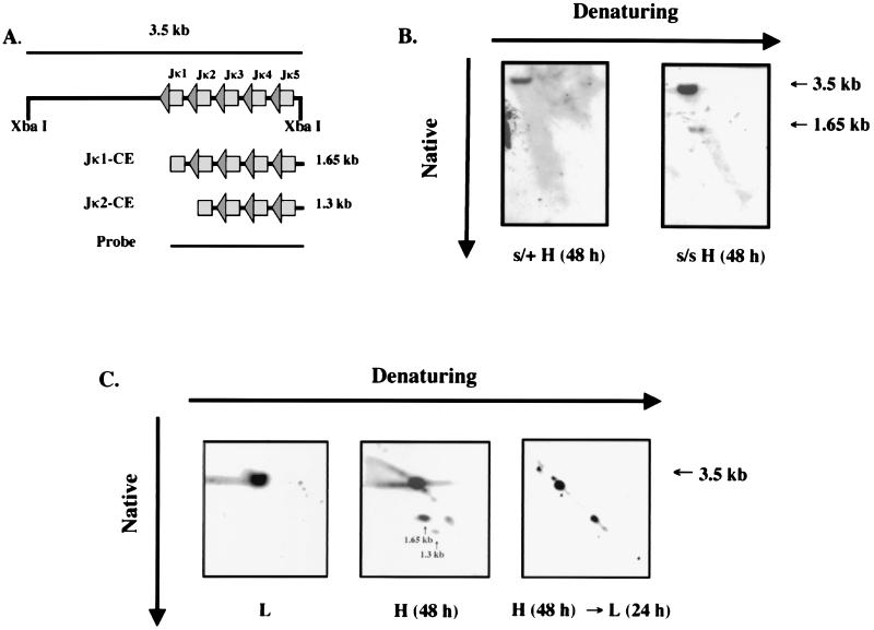
Figure 4.
Hairpin-coding end detection by 2-D electrophoresis. (A) Diagrammatic representation of Jκ-gene rearrangement showing the predicted coding ends, the germline fragment after XbaI digestion, and the probe used for detection. (B) 2-D gel analysis was conducted on the DNA samples prepared from both s/+-ts cells and scid-ts cells cultured at 39°C for 48 hr. The XbaI-digested DNA were electrophoresed in the native condition for the first dimension and in alkaline condition for the second dimension. The germline Jκ fragment as well as the ends are indicated by the arrows. (C) 2-D gel analysis was conducted on the DNA samples prepared from the following cells: scid-ts cells cultured at 33°C (Left), scid-ts cultured at 39°C for 48 hr (Center), and scid-ts cultured at 39°C for 48 hr followed by an incubation at 33°C for 24 hr (Right). Expected sizes are 3.5 kb for the germline κ-locus, 1.65 kb for the Jκ-1 coding ends, and 1.3 kb for the Jκ-2 coding ends, as indicated by the arrows.

