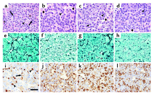
Figure 2.
Histological analysis of anterior pituitaries from 14-month-old female mice. Top row shows hematoxylin and eosin–stained pituitary sections of wild-type (a), Drd2–/– (b), Prlr–/– (c), and compound mutant (Drd2–/–, Prlr–/–) (d) mice. The wild-type pituitary has a normal architecture and mixture of cells including large acidophilic somatotrophs (asterisks), basophils with juxtanuclear, complex lysosomal bodies (arrows), and scattered chromophobic lactotrophs with clear, juxtanuclear Golgi regions (arrowheads). The three mutant genotypes (b–d) all exhibit peliosis (extravasated erythrocytes not contained in capillaries; diamonds) and hypertrophic lactotrophs with very large Golgi regions (arrowheads) and scattered, large, hyperchromatic, atypical nuclei. Middle row: Gordon-Sweet silver stain shows the normal acinar pattern and reticulin network (arrows) in the wild-type pituitary (e), but reveals significant disruption of the reticulin network in Drd2–/– mice (f) and expanded acini and partial loss of reticulin in the Prlr–/– (g) and compound mutant (Drd2–/–, Prlr–/–) (h) mice, findings that are pathognomonic for adenomatous transformation of pituitary cells. Bottom row: PRL immunohistochemistry. The wild-type pituitary (i) contains many mammosomatotrophs with pale, diffuse immunostaining (arrow) and a few mature active lactotrophs with PRL immunostaining in the Golgi apparatus (arrowheads). Almost all the cells in the Drd2–/– (j) and Prlr–/– (k) tumors are active lactotrophs with PRL immunoreactivity in the Golgi. The compound mutant (Drd2–/–, Prlr–/–) (l) tumor is similar, but has fewer immunopositive cells than either single mutant. Representative fields of adenomas from each genotype are shown. Scale bar represents 20 μm; original magnification: ×100, oil immersion.

