Figure 1.
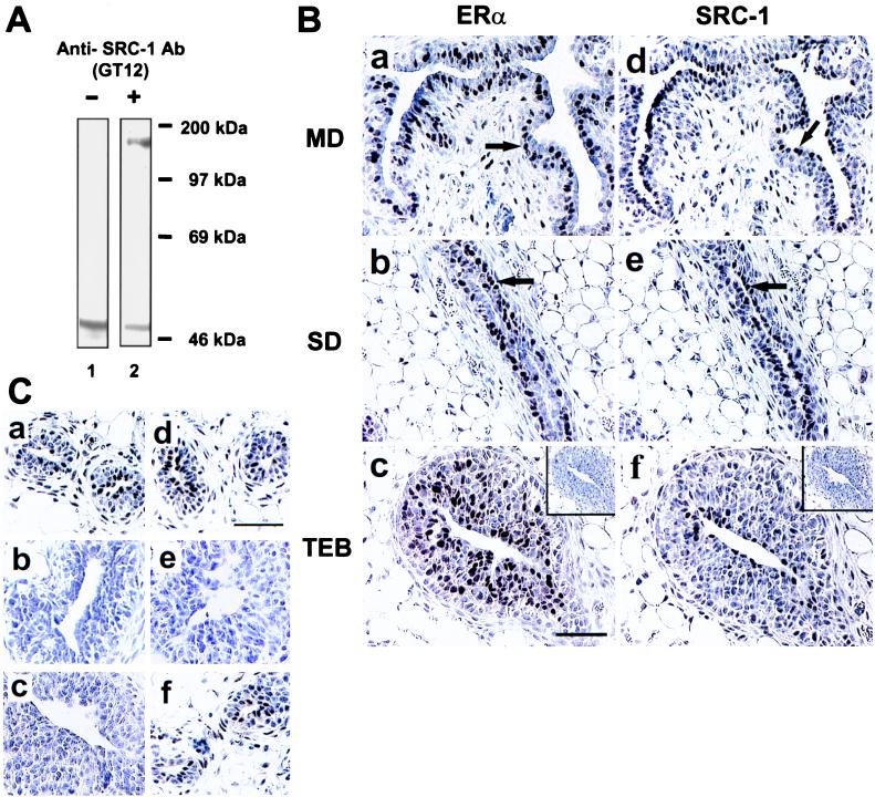
Expression of SRC-1 and ERα in the rat mammary gland. (A) Mouse anti-SRC-1 (GT12) antibody recognized rat SRC-1 (lane 2) as demonstrated by Western blot analysis of GT12 using rat uterine tissue lysate. The endogenous rat IgG from tissues was detected by the secondary antibody, and this signal was also detected without primary antibody (lane 1). (B) Immunohistochemical staining of ERα (a–c) and SRC-1 (d–f) on adjacent sections from main ducts (MD), small ducts (SD), and terminal end buds (TEB) of the mammary gland from 3-week-old virgin female rats. Control specimens stained without primary antibodies are shown in c and f (Insets). The arrows are pointing to ERα-positive cells that are found in a layer closer to the basement membrane and SRC-1-positive cells that exist in a more luminal layer. (C) Immunoreactive SRC-1 was detected by both anti-SRC-1 antibodies, GT12 (a) and M-20 (d), in rat mammary gland. A control specimen (c) stained without primary antibody showed no staining signal. Preabsorption of GT12 or M-20 antibodies with SRC-1 fusion protein (b) or M-20-specific peptide (e), respectively, out-competed the staining signal. M-20 peptide did not diminish the staining signal detected by GT12 (f). Sections were counterstained with Harris hematoxylin. (Bars = 50 μm in B and C.)

