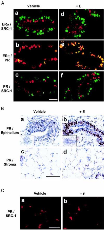
Figure 3.

Segregation of SRC-1 from both ERα and PR in rat mammary epithelial cells when treated with estrogen. Mammary gland from 3-week-old virgin female rats treated with vehicle (sesame oil) or 1 μg of estrogen benzoate (+E) for 24 h were used. (A) Dual immunofluorescent labeling of ERα/SRC-1, ERα/PR, and PR/SRC-1. (a and d) ERα (green) and SRC-1 (red) were stained simultaneously with MC-20 and GT12. (b and e) ERα (red) and PR (green) were stained simultaneously with MC-20 and anti-PR IgG (MA1–410). (c and f) PR (green) and SRC-1 (red) were stained simultaneously with MA1–410 and M-20. (Bar = 100 μm.) (B) Expression of PR in both epithelium and stroma of the mammary glands. Control specimens stained without primary antibody are shown in insets. (Bar = 50 μm.) (C) Dual immunofluorescent labeling of PR and SRC-1 in stroma of the mammary glands. The staining was performed as in A. (Bar = 100 μm.)
