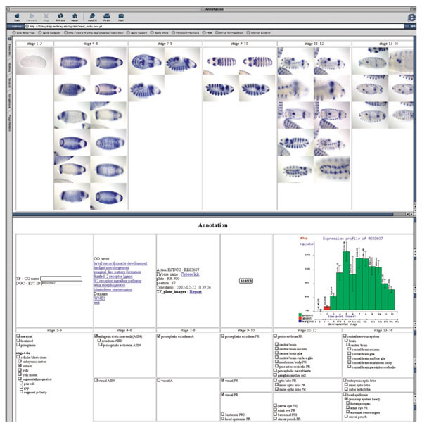
Figure 2.

Imaging expression patterns during embryogenesis. Screen shot of the web-based tool used to organize and annotate captured images. The figure shows the categorization of images in six stage ranges, reflecting developmental time, presented left to right. The annotation terms are similarly arranged from left to right and grouped from top to bottom according to related organ systems. Groups of images at a given stage range will be associated with groups of annotation terms appropriate for that stage range. Microarray profiles and links to public databases are also included.
