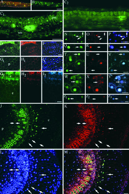
Figure 3.
Induction of BrdU Incorporation by TGMV Is Cell Autonomous.
Systemically infected tissues were incubated with 100 μM BrdU for 24 h as described for Figure 1, fixed, and Vibratome sectioned. BrdU was detected using anti-BrdU antibodies and Alexa 488–conjugated secondary antibodies (green in [A] to [D], [G1], [G2], [J], [M], [N], [Q], [T], [W], and [Z1]). Sections also were hybridized with Texas Red–labeled oligonucleotide probes to detect TGMV DNA (red in [E], [H1], [H2], [K], [M], [O], [R], [U], [X], and [Z2]) and stained with DAPI (blue in [A], [F], [I1], [L], [M], [P], and [S]). TGMV AL1 was immunolocalized with anti-AL1 antibodies and Marina Blue–labeled secondary antibodies (blue in [I2], [V], [Y], and [Z3]).
(A) and (B) BrdU incorporation (green) in an immature healthy leaf is confined to isolated nuclei throughout the lamina. A triple-wavelength excitation filter was used in (A) to show BrdU-incorporating nuclei among nuclei with no BrdU (blue).
(C1) BrdU incorporation into nuclei of a TGMV-infected mature leaf. Nuclei of adjacent palisade mesophyll cells and some spongy mesophyll cells show abundant BrdU labeling.
(C2) Mock-inoculated control mature leaf section. Bright autofluorescence of vascular tissue is visible.
(D) to (F) TGMV-infected root section. Viral DNA and BrdU are evident in cortical cells.
(G1) to (I2) Mock-inoculated control stem cross-sections. Double nuclei are common in N. benthamiana pith. Low levels of background BrdU incorporation are observed in some nuclei.
(J) to (M) TGMV-infected stem cross-section. BrdU (green) and viral DNA (red) are colocalized. (M) shows a three-color overlay of (J) to (L), demonstrating colocalization of BrdU and viral DNA in DAPI-stained nuclei. Single arrows show nuclei with low amounts of viral DNA and correspondingly lower BrdU signals. Nuclei between double arrows lack viral DNA and BrdU.
(N) to (S) TGMV-infected stem sections. Arrows show nuclei that have high BrdU but minimally detectable viral DNA. Blue indicates DAPI.
(T) to (Z3) TGMV-infected stem sections. Arrows show nuclei that have BrdU and minimally detectable viral DNA but contain TGMV AL1. Blue indicates AL1-specific signal.
c, cortex; e, epidermis; pm, palisade mesophyll; sm, spongy mesophyll; v, vascular tissue. Bars = 100 μm for (A) to (M), (V), and (Z3) and 50 μm for (P), (S), and (Y).

