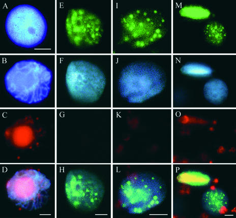
Figure 6.
Host Chromatin of TGMV-Infected Nuclei Is Distributed Evenly and Resembles S-Phase Chromatin Organization.
Stem tissues were pulse labeled with BrdU for 4 h and then processed and imaged as described for Figure 5. For (E) to (P), BrdU (green) is shown in the top row, DAPI-stained host chromatin (blue) is shown in the second row, TGMV DNA (red) is shown in the third row, and three-color overlays are shown in the fourth row except for (D), which was imaged using a triple cube.
(A) Healthy interphase nucleus stained with DAPI showing typical dispersed chromatin.
(B) to (D) Advanced-stage TGMV-infected nucleus showing high viral DNA accumulation and condensed chromatin.
(E) to (L) TGMV-infected nuclei showing BrdU foci and no detectable viral DNA. Host DNA in these nuclei is dispersed evenly.
(M) to (P) A nucleus with BrdU foci but no detectable viral DNA adjacent to a nucleus with viral DNA and uniform BrdU label.
Red objects outside of the nuclei in (C), (K), (L), (O), and (P) are autofluorescent chloroplasts. Bars = 5 μm.

