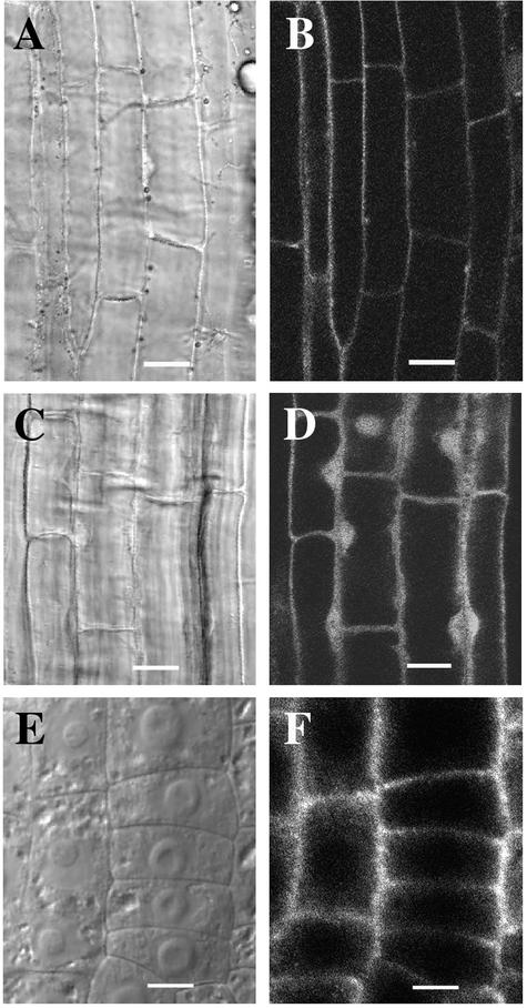
Figure 9.
Localization of FRA1 in Root Cells.
Roots of 3-day-old seedlings expressing the FRA1-GFP fusion protein ([A] and [B]) or GFP alone ([C] and [D]) were used to visualize GFP signals. Roots of 3-day-old wild-type seedlings were used for the immunolocalization of FRA1 ([E] and [F]).
(A) and (B) Differential interference contrast (DIC) image (A) of root cells and corresponding fluorescence signals of FRA1-GFP (B). The signal was present in the cytoplasm but absent in the nucleus.
(C) and (D) DIC image (C) of root cells and corresponding fluorescence signals of GFP (D). The signal was present in both the cytoplasm and the nucleus.
(E) and (F) DIC image (E) of root cells and corresponding FRA1 protein localization (F). FRA1 was present in the cytoplasm but absent in the nucleus. Note that the DIC image (E) shows the prominent nuclei in the centers of the cells.
Bars = 12 μm in (A) to (D) and 6 μm in (E) and (F).

