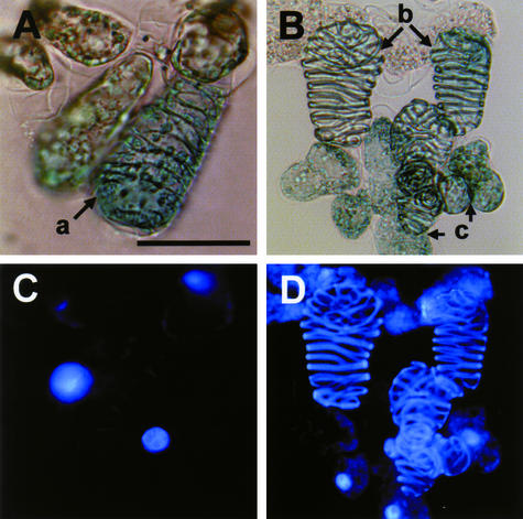
Figure 5.
Light and Fluorescence Images of Zinnia Cells Stained with X-Gluc Solution and 4′,6-Diamidino-2-Phenylindole.
p35SZEN1A and p35SGUS were introduced by particle bombardment into Zinnia cells cultured for 40 h in TE-inductive D medium. Collected cells were fixed and then incubated with X-Gluc solution (see Methods) for 12 h. 4′,6-Diamidino-2-phenylindole staining was performed for 15 min in the dark. The transformed cells, which were identified by the expression of GUS activity (blue staining), were categorized into three types by cell shape and staining pattern of 4′,6-diamidino-2-phenylindole: GUS-positive TEs with the nucleus (a); GUS-positive TEs without the nucleus (b); and GUS-positive non-TE cells with the nucleus (c). (A) and (C) show light images, and (B) and (D) show fluorescence images. Bar = 50 μm.

