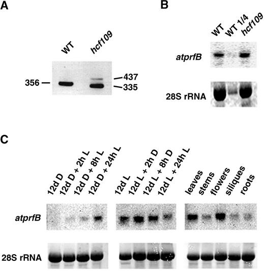
Figure 2.
RT-PCR and RNA Gel Blot Analysis of atprfB.
(A) Specific primers of exon 1 and exon 3 (ex1-f and ex3-r, respectively) were used for RT-PCR of mutant and wild-type leaf mRNA. DNA fragments of the indicated sizes (bp) were separated on an agarose gel and visualized with ethidium bromide staining. The top band in the mutant lane represents the unspliced form of atprfB, and the bottom band represents the incorrectly spliced form of atprfB. WT, wild type.
(B) Expression levels of atprfB from 3-week-old mutant and wild-type leaves of plants grown under identical conditions and analyzed by RNA gel blot hybridization. Levels of 28S rRNA are shown to demonstrate uniform loading.
(C) The effect of light (L) and dark (D) incubation on the expression of atprfB in seedlings and the levels of atprfB in different tissues were analyzed in the wild type using RNA gel blots, as described in Methods.

