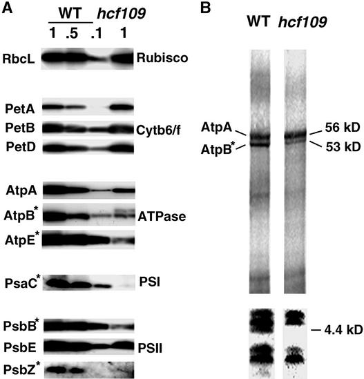
Figure 6.
Accumulation and Labeling of Plastid Proteins.
(A) Immunoblot analysis of thylakoid membrane proteins from 3-week- old hcf109 and wild-type seedlings. For quantification, 100, 50, and 10% of wild-type membranes were loaded. Asterisks indicate proteins encoded by genes containing a TGA stop codon. Cytb6f, cytochrome b6/f; PSI and PSII, photosystems I and II; Rubisco, ribulose-1,5-bisphosphate carboxylase/oxygenase; WT, wild type.
(B) In vivo labeling of chloroplast membrane proteins separated on 10% (top) and 16% (bottom) polyacrylamide gels. Coseparation of molecular mass standards served to estimate the sizes (in kD) of the labeled proteins.

