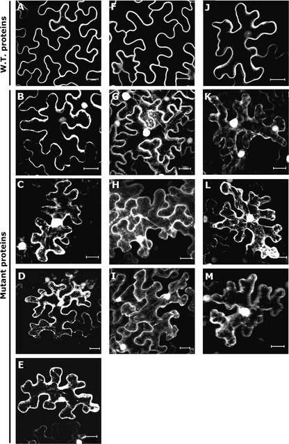
Figure 4.
Effect of C-Terminal Cys Residues on the Association of AtRAC7, AtRAC8, and AtRAC10 with Membranes.
Wild-type and mutant GFP-AtRACs were expressed transiently in N. benthamiana leaf epidermal cells.
(A) to (E) AtRAC7.
(F) to (I) AtRAC8.
(J) to (M) AtRAC10.
(A), (F), and (J) show wild-type proteins. Cys mutants were designated with the suffix mS and received a number specifying the amino acid position. These are shown as follows: Atrac7mS206 (B); Atrac7mS203 (C); Atrac7mSS203+206 (D); Atrac7mS196 (E); Atrac8mS205 (G); Atrac8mS199 (H); Atrac8mSS199+205 (I); Atrac10mS208 (K); Atrac10mS202 (L); and Atrac10mSS202+208 (M). Nuclei and cytoplasmic strands were visualized in the Cys mutants, indicating that the association of fusion proteins with the plasma membrane was weaker. Note that with the exception of Atrac7mS206, in which dissociation from the plasma membrane was weaker, mutations in a single Cys were similar in effect to mutations in two Cys residues. All images are from projection stacks of multiple confocal sections taken with a confocal laser scanning microscope. Bars = 20 μm.

