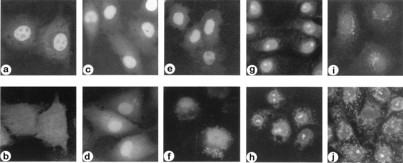
Figure 2.
Subcellular localization of fluorescein-labeled nido- and closo-OPDs in vitro. (a and b) The same set of cells 2 h after coinjection of nido-(CB)5 (5 μM solution) and rhodamine-labeled BSA (1 mg/ml) into the cytoplasm. When viewed at their characteristic UV wavelengths, nido-(CB)5 appears in the nucleus (a) and BSA appears in the cytoplasm (b). (c and d) Rapid nuclear accumulation of nido-(CB)5 (100 μM solution) (c) and nido-(G1)5 (100 μM solution) (d) within 10 min after cytoplasmic microinjection. (e and f) Long-term nuclear retention of nido-(CB)5 (100 μM solution) (e) and nido-(G1)5 (100 μM solution) (f) in the cells 24 h after cytoplasmic microinjection. (g and h) Distribution of OPD in subcellular compartments in digitonin permeabilized cells incubated with nido-(CB)5 (g) and nido-(G1)5 (h). (i and j) Subcellular distribution of closo-(G1)5 in the cells 2 h after microinjection (100 μM solution) (i) and in digitonin permeabilized cells (j).

