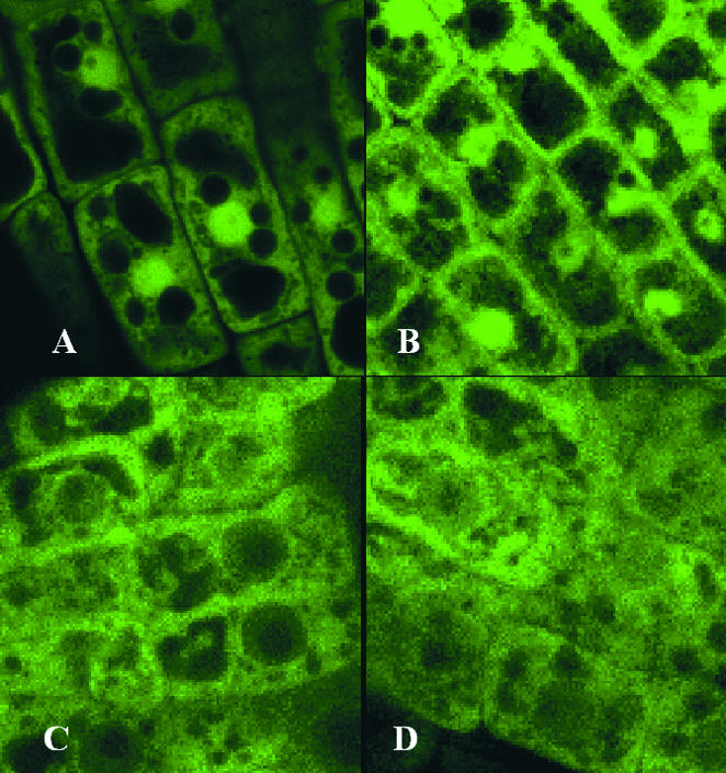
Figure 4.

14-3-3–GFP Fusions Show Diverse Localization Patterns.
(A) GFP is localized to several areas of the Arabidopsis root cells and tends to concentrate to some extent within the nuclei.
(B) to (D) Fusion proteins in which GFP is located at the C terminus of diverse 14-3-3s tend to localize differentially within Arabidopsis root cells. The GFP κ signal is relocalized prominently to the plasma membrane region but remains concentrated in the nucleus (B). The GFP υ signal is largely driven out of the nucleus and into the cytoplasmic compartments (C), whereas the GFP φ signal is present rather diffusely throughout (D). Coding regions for the 14-3-3s were amplified by polymerase chain reaction, placed as translational fusions with GFP in pBI12135sGFP(S65T), and transformed into Arabidopsis Wassilewskija by vacuum infiltration. Five-day-old plants were visualized using an Olympus IX70 inverted microscope (Tokyo, Japan) mounted to a Bio-Rad MC 1024ES laser scanning system with 24-bit confocal imaging. Images were Kalman averaged four times.
