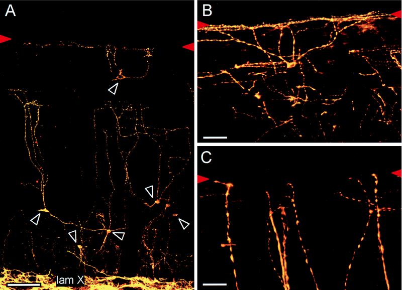
Figure 1.
(A) NK-1 receptor immunoreactive neurons (white arrowheads) are scattered through this 30-μm midsagittal section of the lumbar spinal cord of the adult rat. The dense NK-1 receptor immunoreactivity in lamina X (lam X) distinguishes the gray matter from the white matter of the DCs. (B and C) The neurons are also MAP-2 positive and have highly varicose dendrites that arborize extensively, often contacting the dorsal surface of the cord (red arrowheads). (Bars: 100 μm, A; 50 μm, B; and 25 μm, C.)

