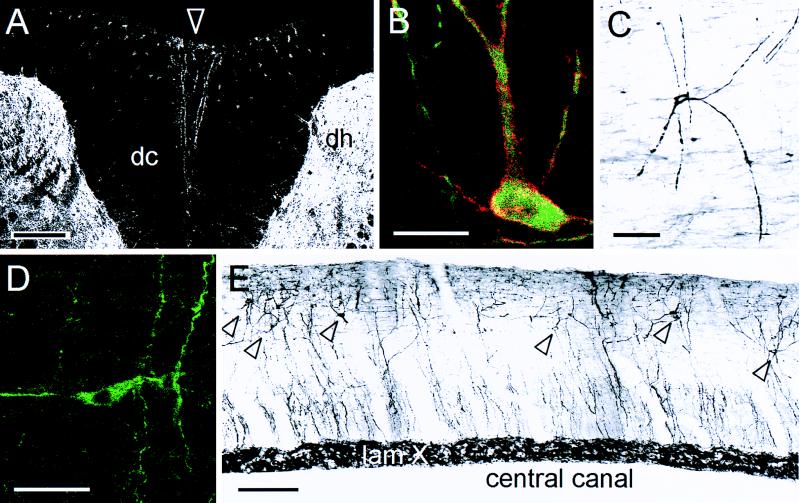
Figure 2.
(A) Although the neurons in the DCs (dc) are best viewed in sagittal section, MAP-2 immunoreactive dendrites occasionally can be seen along the midline (arrowhead) in transverse sections. The dendrites are located at a distance from the heavily labeled dorsal horn gray matter (dh). (B) Superimposition of confocal images for MAP-2 (green) and the NK-1 receptor (red) reveals that every DC neuron labels for both molecules. Where there is overlap, the image appears yellow. C and D illustrate DC neurons from midsagittal sections of the cervical spinal cord in monkey and bird (Zebra finch), respectively. Both are stained for MAP-2 by using an immunoperoxidase (C) or an immunofluorescence protocol (D). (E) This midsagittal montage of sections from a 13-day-old rat spinal cord illustrates the widespread distribution of the DC neurons (labeled with MAP-2; arrowheads); many are located close to the surface of the cord, distant from the gray matter (lamina X) around the central canal. A, B, and D are confocal images; C and E were digitized from slides and prepared with Adobe Photoshop. (Bars: 200 μm, A; 20 μm, B; 50 μm, C; 4 μm, D; and 50 μm, E.)

